Diagnostics
Overground (“telemetric”) Endoscopy has revolutionised what we know about the upper respiratory tract of horses and how diseases can affect performance. We use the finest and latest technology on the market to get crystal clear images of your horse’s throat during exercise, allowing us to diagnose subtle pathology that can severely limit performance.
When a horse is heard to make a noise during exercise the initial step would be to use a conventional endoscope (a long flexible camera) to examine the horse’s upper respiratory tract (throat and airway). Some conditions can be diagnosed at rest but others only occur during exercise. Overground endoscopy (OGE) is an advanced technique that allows us to examine the upper respiratory tract when a horse is galloping. The technique is also useful in horses that are not running or working as well as expected.
An endoscope is inserted up the nostril and positioned to focus on the larynx at the back of the throat. The camera is held in place on a specially designed bridle and is connected to a computer system mounted beneath the saddle. This records the video and also sends a live feed back to a wireless handheld monitor so that we can look at the horse’s larynx as it is exercised. The horse is then exercised normally, but at a good pace to exacerbate any issues, and we follow in a vehicle with the monitor.
Once we know what the problem is we can discuss options with connections and a few different surgeons and work out the best way forward to get the horse back to peak performance.
Where the overground is performed is dependent on the horse and the issue. For racehorses we would generally come to your normal gallop and for sports and pleasure horses we would generally come to your yard.
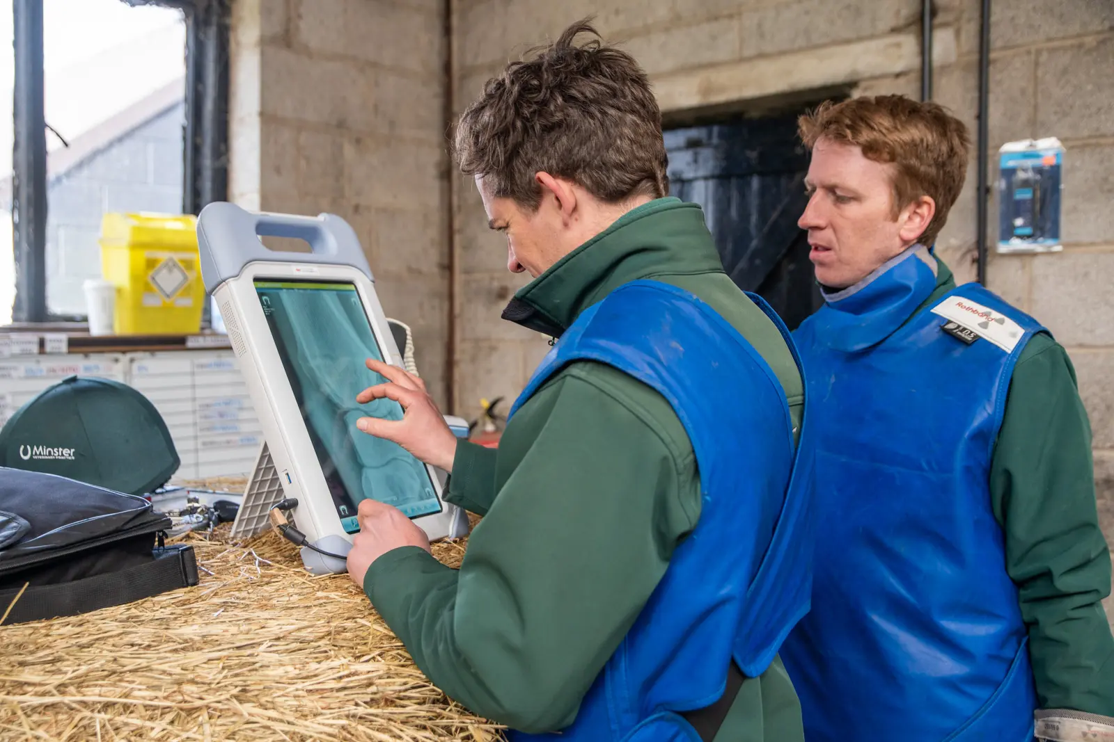
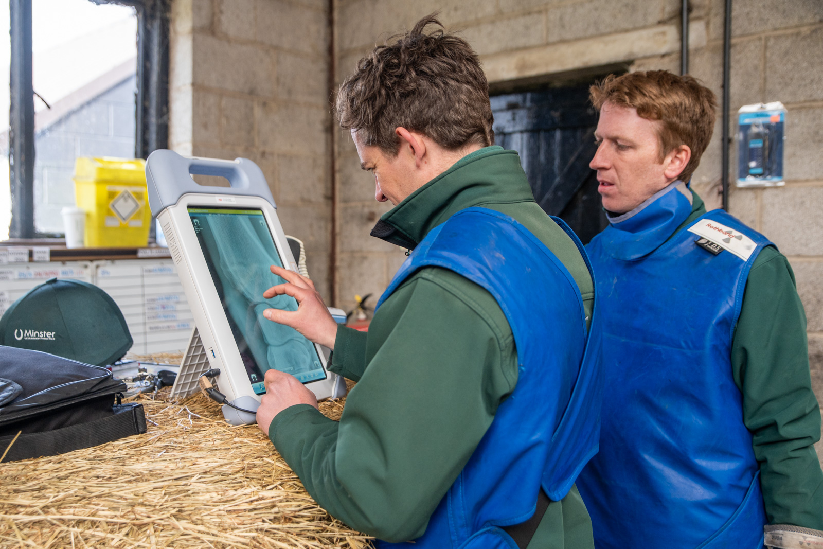
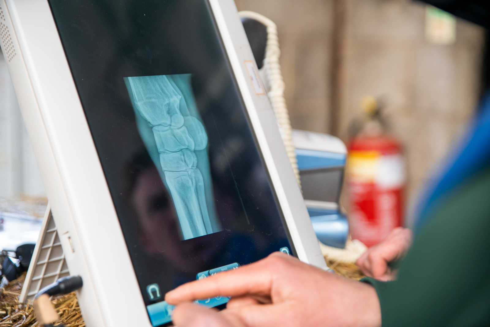
X-rays are a vital part of most lameness investigations, and we are very lucky to have three state of the art, portable Cuattro Slate DR x-ray systems. These digital radiography machines are a huge asset to the practice.
They, along with the generators are completely wireless, so are incredibly useful for our ambulatory vets who can take x-rays with ease out on yards. Not only do we have improved safety as there are no trailing wires, the machines are incredibly fast and provide extremely high quality diagnostic images.
We are also able to use them in remote locations with no electricity, allowing us to provide a better service to all of our clients.
Our ultrasound scanners can be used by our experienced team of vets to perform ultrasound of the limbs, reproductive ultrasound, and abdominal ultrasound to diagnose a range of issues. Our scanners include portable options that can be used at your yard or at our clinics, to ensure that your horse receives a timely diagnosis and treatment plan.
What is ultrasonography?
Ultrasonography is a valuable and widely used diagnostic tool in horses, which diagnose and evaluate a number of conditions. An ultrasound captures live images from inside the horse’s body and unlike some other imaging techniques, it does not use radiation.
Advantages of ultrasonography
- Non invasive process
- Painless
- Ultrasound images are real-time, so you are able to see direct visualisation
- Ultrasonography does not use radiation
- Doppler ultrasound can measure the speed of blood flow
- Ultrasound captures images of soft tissue that don’t show up on an x-ray
- The equiment is mobile
- Ultrasounds are usually quick, with most sessions lasting around 20-30 minutes
How does it work?
Ultrasound uses high-frequency sound waves that pass through the body before reflecting and bouncing back to the handpiece. This echo is transformed into a picture by the computer. Different tissues reflect the ultrasound in different ways and this allows us to visualise structures in great detail. Additionally, colour doppler ultrasound uses the way ultrasound reflects differently in moving blood to show how it is flowing.
What can be seen?
Musculoskeletal system
Ultrasonography is used extensively in lameness investigations for the scanning of tendon and ligament injuries as well as assessing wounds, joint surfaces, fractures and soft tissue swellings. Ultrasonography may also be useful for the detection of back and pelvic injuries.
Heart and vascular system
Ultrasonography of the heart is important to assess the chambers and valves of the heart and is invaluable in the assessment of the significance of many types of heart murmur. Colour flow Doppler is used to assess dynamic blood flow through different parts of the heart. Ultrasonography may also be useful for assessing thrombi and peripheral blood vessels.
Abdomen
Ultrasonographic examination of the intestines and other abdominal structures (eg. liver, kidney and spleen) is an important diagnostic tool in the investigation of horses with colic, weight loss or diarrhoea. Ultrasound guidance is frequently used to allow safe and precise biopsy of internal structures such as the liver, lungs and kidneys.
Reproductive Tract
Ultrasonographic assessment of the ovaries and uterus is important in the management of brood mares to assess the reproductive tract, stage of oestrous cycle and pregnancy diagnosis. Regular ultrasound scans of the ovaries are performed (every few hours) of mares undergoing artificial insemination (AI) to ensure that insemination is performed at the optimal time. Early pregnancy diagnosis is important to ensure that a mare is not carrying twins.
Thorax
Ultrasonographic assessment of the thoracic cavity, including the lungs, is important in the assessment of horses with pneumonia, lung masses or pleurisy.
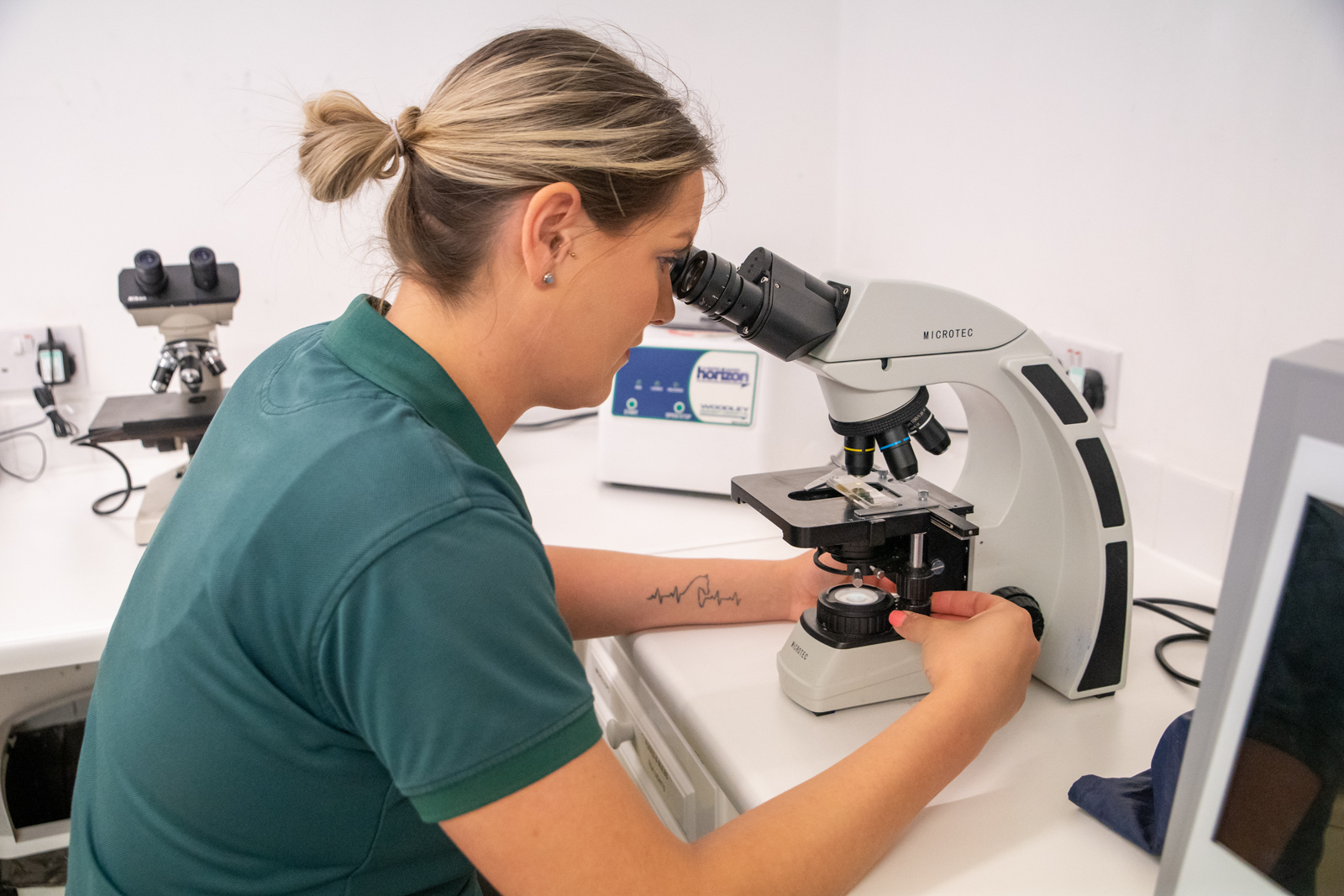
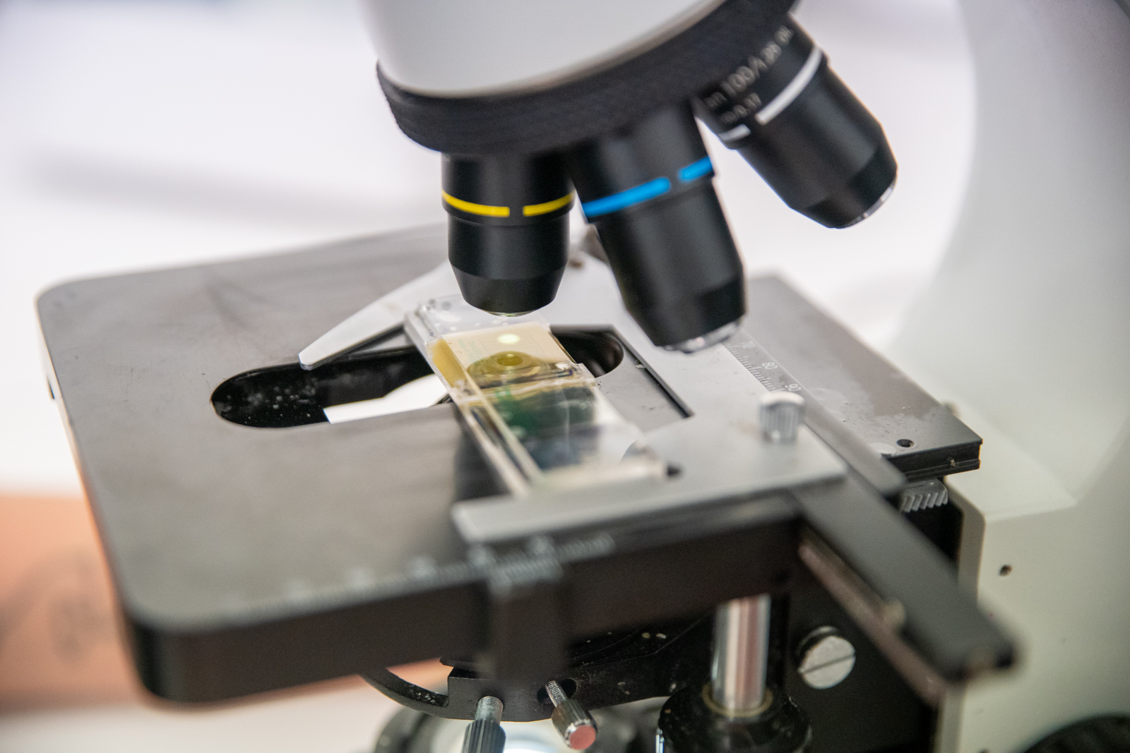
At Minster Vets we are able to provide a full range of blood tests for a variety of conditions. We have our own in-house laboratory in which we can perform general haematology and biochemistry blood profiles, providing same day results. We also have multiple readers for measuring serum amyloid A, which can provide stable side results in just 10 minutes. We have good working relationships with multiple other laboratories across the UK which means all tests can be catered for, including but by no means limited to strangles serology, Cushing’s (ACTH) and equine metabolic syndrome, foal iGg, pre-breeding and export bloods.
Haematology
Our haematology profile assesses your horse’s red and white blood cells, as well as total protein levels and fibrinogen. We use this most commonly to check for signs of dehydration, inflammation or anaemia, but it can also help to support a diagnosis of bacterial or viral infection.
Biochemistry
A biochemistry profile provides an assessment of proteins, enzymes, glucose, and various metabolites. Specific combinations of abnormalities can give an indication of particular body systems which may be affected, such as the liver, kidneys or muscles, and results can also provide support for diagnoses of intestinal or metabolic abnormalities. A biochemistry profile is extremely useful in the diagnosis and monitoring of many conditions and is particularly useful in the work up of medical cases.
Serum Amyloid A
SAA is a protein produced in the acute stages of inflammation and infection. It is an invaluable tool, not only in the diagnosis of inflammatory conditions, but also in monitoring your horse’s response to treatment, as the value will fall rapidly once appropriate treatment is instigated. It allows us to make more informed choices about our treatment plans, and in particular the use of antibiotics. It is an extremely rapid test, providing a result in just 10 mins and is a small portable machine, making it extremely popular in our racing and stud yards.


